02 / RIRS · flexible ureterorenoscope · laser
RIRS and flexible endoscopy for kidney stones in Krasnoyarsk
RIRS uses a flexible endoscope passed through the urethra and ureter to reach the renal calyces, without an external incision. The stone is fragmented with a laser; the strategy depends on its burden, location and kidney anatomy.
- RIRS
- Flexible endoscopy
- Laser
This material was prepared by a doctor using Russian clinical guidelines and EAU guidance. It does not establish a diagnosis, prescribe treatment or replace an in-person consultation. The choice of method depends on investigations, coexisting conditions and individual risks. A surgical outcome cannot be guaranteed in advance.
- Route
- through the natural urinary tract
- Optics
- flexible ureterorenoscope
- Important
- large stones may require staged treatment
01 / Indications
When RIRS may be an option
- kidney stones up to 20 mm
- several small stones or a difficult location in the calyces
- unsuccessful shock wave lithotripsy
- situations where percutaneous access is undesirable
- selected cases with stones larger than 20 mm, with an understanding that several stages may be needed
02 / Decision
What affects the choice of access
The doctor assesses total stone volume and density, the position of the lower calyx, ureteral width and anatomy, kidney function, infection, previous operations and previously placed stents.
For large stones, RIRS is compared with percutaneous access. The goal is not to avoid an incision at any cost, but to choose a method with a reasonable balance of effectiveness, number of stages and risks.
03 / Surgery
How RIRS is performed
A flexible ureterorenoscope is passed through the urethra, bladder and ureter into the kidney's collecting system. The stone is fragmented with a laser or individual pieces are retrieved with a basket under direct vision.
A temporary ureteric stent may be inserted at the end. If the ureter is narrow, it may be safer to place a stent first and perform the main procedure after passive dilation.
Clinical practice
Flexible endoscopy in clinical practice
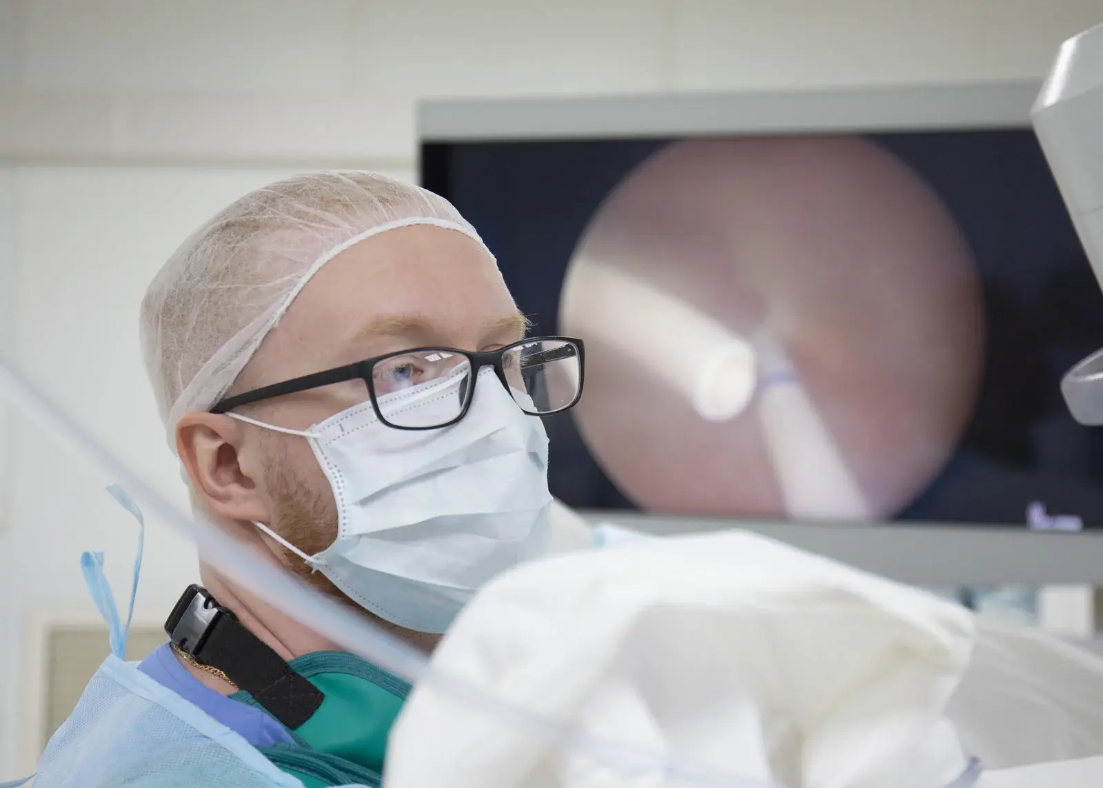
The endoscopic images show how a stone is visualised and fragmented inside the kidney's collecting system. They cannot determine the treatment method without CT, laboratory tests and an in-person assessment.

Working with a flexible endoscope through the natural urinary tract under continuous visual control
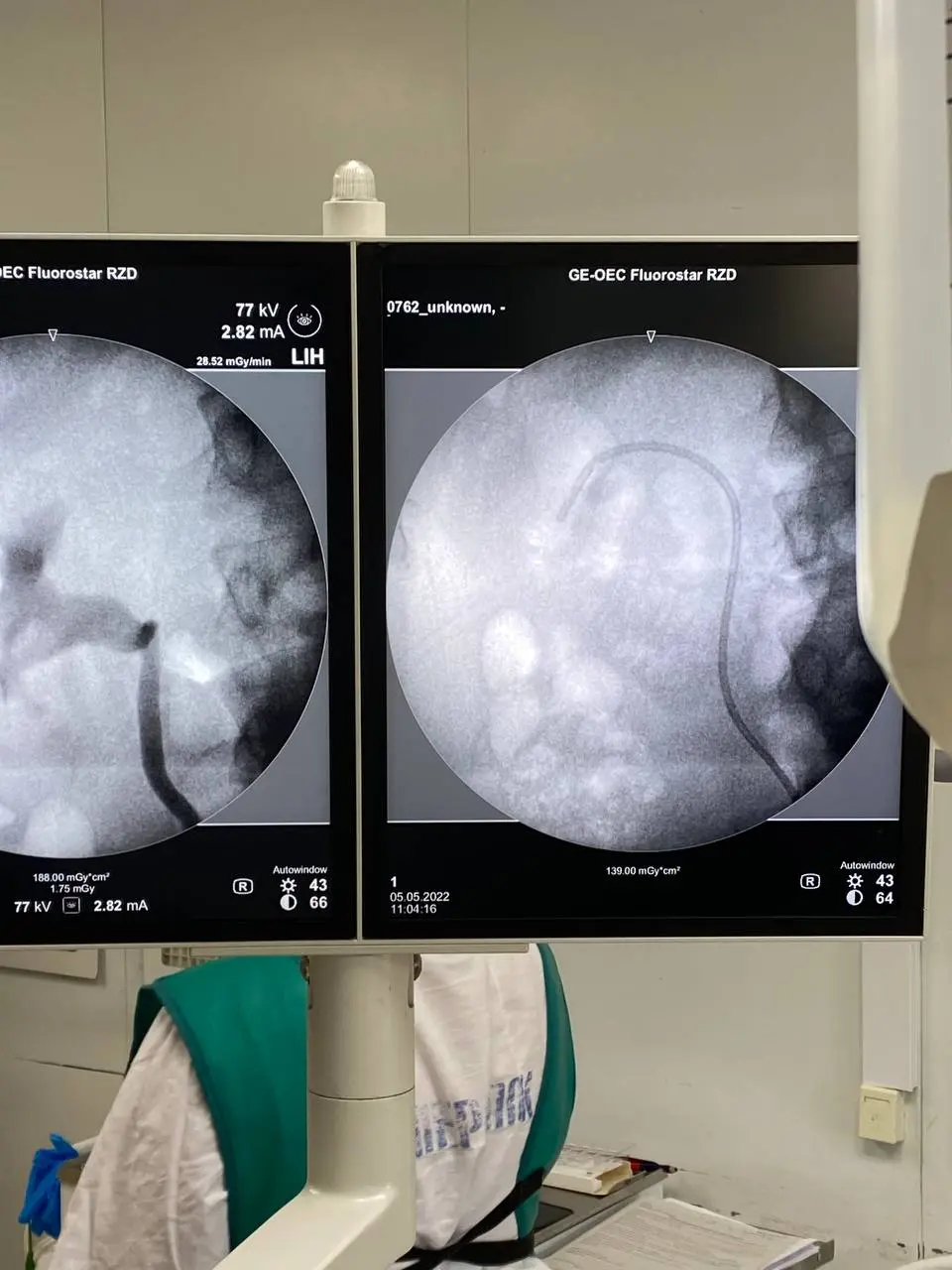
X-ray verification of guidewire and instrument position during access to the renal collecting system
Retrograde intrarenal surgery
An RIRS excerpt: reaching a stone through the natural urinary tract and working with a flexible endoscope. Video without sound; the extent and staging of treatment are determined individually.
Flexible endoscopy of the renal calyces
A clinical excerpt of retrograde intrarenal surgery. The video shows a single stage and cannot determine the treatment method without CT, laboratory tests and an in-person assessment.
04 / Preparation
What to prepare for the appointment
- CT in DICOM format and the report
- urinalysis and urine culture
- creatinine and other kidney function results
- tests for anaesthesia and coagulation assessment
- information about previous procedures and stents
- a list of medicines and allergies
05 / After surgery
Stent, symptoms and follow-up
In the first few days, a little blood, frequent urgency and discomfort may occur, especially with a stent in place. The patient should receive a clear plan and removal date in advance.
Follow-up imaging helps assess residual fragments. After removal, stone composition analysis and prevention of recurrence are discussed.
06 / Risks
What complications are possible?
- infection and fever
- bleeding
- ureteral injury or later narrowing
- migration and residual fragments
- stent-related symptoms
- need for another treatment stage
07 / Urgent care
Do not wait for a routine appointment
Fever with chills, inability to urinate, increasing pain, severe weakness or significant blood with clots after a procedure require urgent medical assessment.
Patient questions
Frequently asked questions
Is there an incision after RIRS?
There is usually no external incision: the instrument passes through the natural urinary tract. This does not eliminate internal risks or the need for a temporary stent.
Why a large stone is sometimes treated in several stages
Safe operating time, the amount of fragmentation and the ability to remove fragments are limited. Staged treatment can sometimes reduce the risk of infection and injury.
Is a stent mandatory after RIRS?
Not always. The decision depends on ureteric swelling or injury, infection, residual fragments and difficulty of access.
Verifiable evidence
Sources
This page explains general guidance in plain language. Decisions for an individual patient are made after an in-person assessment and cannot be reduced to a single guideline recommendation.
In-person assessment and a clear plan