05 / PSA · MRI · pathology
Ultrasound- and MRI-guided prostate biopsy in Krasnoyarsk
A biopsy confirms or rules out clinically significant cancer using tissue samples. It is not ordered automatically on the basis of one elevated PSA value: PSA trends, MRI, PSA density, the examination and individual risk factors are assessed first.
- PSA
- mpMRI
- Morphology

This material was prepared by a doctor using Russian clinical guidelines and EAU guidance. It does not establish a diagnosis, prescribe treatment or replace an in-person consultation. The choice of method depends on investigations, coexisting conditions and individual risks. A surgical outcome cannot be guaranteed in advance.
- Goal
- obtain tissue samples for histopathological examination
- Before biopsy
- MRI and overall risk assessment
- Guidance
- Ultrasound informed by MRI findings
01 / Indications
When a biopsy may be needed
- suspicious MRI findings
- elevated or rising PSA after possible causes have been reviewed and the test repeated
- suspicious digital rectal examination findings
- persistent risk after a previous negative biopsy
- further assessment of an already identified condition or active surveillance for selected indications
02 / Decision
Why PSA alone is not enough
The decision considers PI-RADS, PSA density and changes, family history, age, previous biopsies and whether the result would change further treatment. When localised disease is suspected, MRI is usually performed before biopsy.
A suspicious lesion is sampled with targeted biopsy, with additional regional or systematic sampling when indicated. The specific approach and extent of biopsy are discussed in advance.
03 / Procedure
How tissue samples are obtained
Under ultrasound guidance, the needle takes thin tissue cores from the required prostate regions. If MRI shows a lesion, targeted guidance is used. Samples from different sites are labelled and sent for histopathological examination.
The transperineal approach is usually associated with a lower risk of infectious complications than the transrectal approach. The choice depends on equipment, the clinical situation and the tissue-sampling plan.
Clinical practice
Prostate biopsy in clinical practice
The images show the doctor, biopsy instrument and labelled tissue cores. The biopsy decision and sampling plan are based on PSA, MRI, examination and individual risk considered together.

Preparing for the procedure: guidance, access and the number of samples are planned from the investigation findings
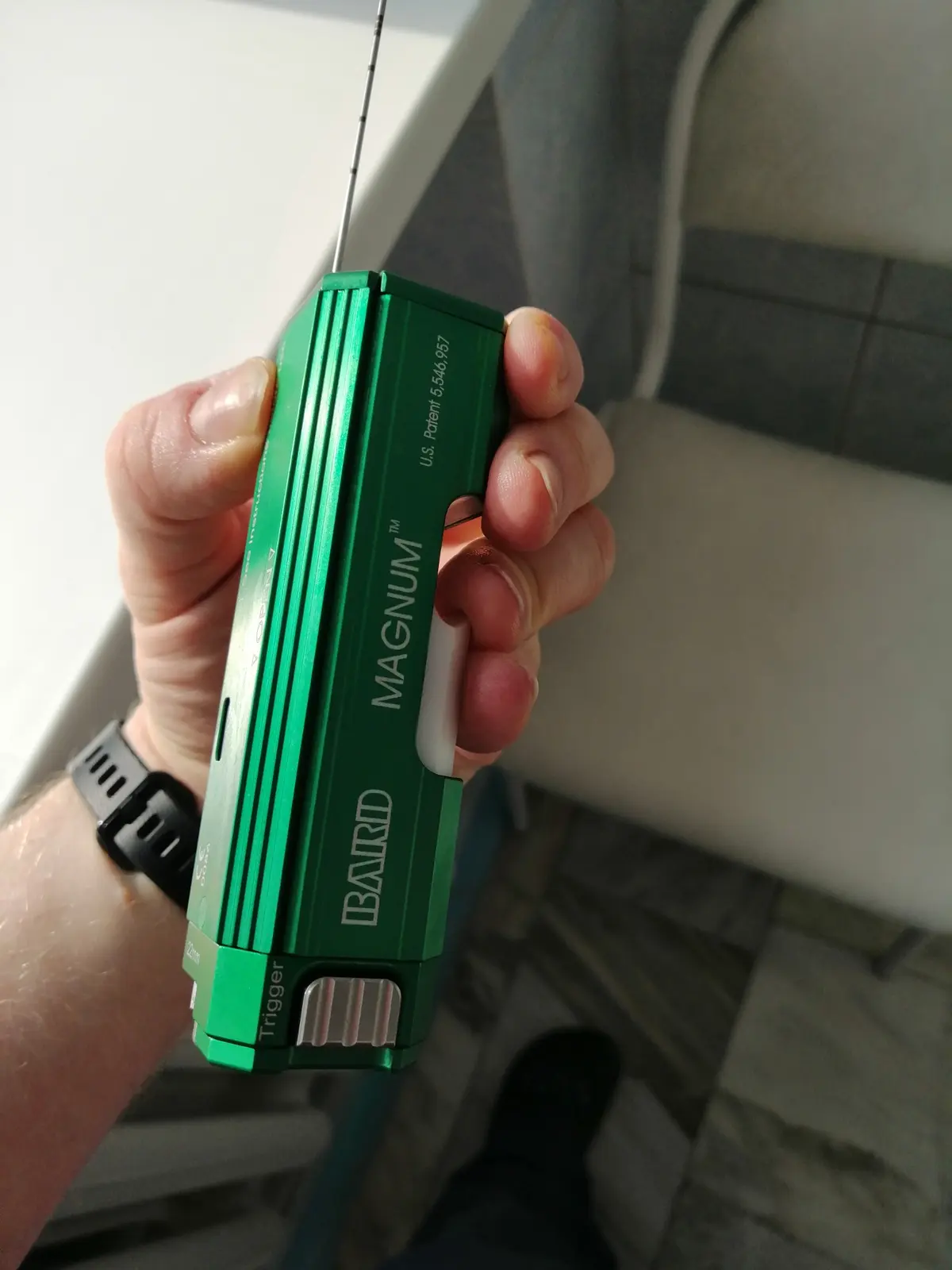
The biopsy instrument obtains thin cores of tissue from predefined areas
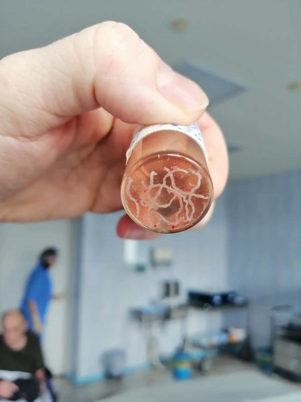
The tissue cores are labeled by area and sent for histopathological examination
04 / Preparation
What to bring and what to discuss
- MRI in DICOM format and the PI-RADS report
- dated PSA results and prostate volume measurements
- previous pathology reports, slides or tissue blocks
- family history of cancer
- blood tests and information about infections
- allergies and medications affecting blood clotting
05 / After the procedure
What may be temporary
A small amount of blood in urine or semen is possible, and with a transrectal approach, bleeding from the rectum may occur. The acceptable duration of symptoms, restrictions and time to receive histology results are explained individually.
A negative biopsy does not always definitively exclude clinically significant disease. Further follow-up depends on the overall risk, MRI and PSA trends.
06 / Risks
Possible complications
- bleeding and pain
- infection, potentially progressing to sepsis
- acute urinary retention
- temporary worsening of urination
- the need for reassessment if risk persists
07 / Urgent care
When to seek immediate care
A high temperature, chills, inability to urinate, severe weakness, increasing bleeding or clots require urgent medical care.
Patient questions
Frequently asked questions
Elevated PSA always means cancer
No. PSA is affected by various conditions and circumstances. Repeat testing, changes over time, prostate volume, MRI, examination and individual risk all matter.
Can a biopsy be done without MRI?
In some situations, yes. However, when localised cancer is suspected, current guidelines generally recommend MRI before biopsy.
What a negative biopsy means
It means that no tumour was found in the samples obtained. If risk persists, monitoring, MRI review or repeat investigation may be needed.
Verifiable evidence
Sources
This page explains general guidance in plain language. Decisions for an individual patient are made after an in-person assessment and cannot be reduced to a single guideline recommendation.
In-person assessment and a clear plan