01 / PCNL · MiniPerc · complex stones
Removal of large and staghorn kidney stones
Percutaneous nephrolithotripsy is endoscopic stone removal through a small access in the lumbar region. It is often considered for large, staghorn and anatomically complex stones; the number of access tracts and stages is determined from CT findings.
- Staghorn stones
- CT planning
- Percutaneous access
This material was prepared by a doctor using Russian clinical guidelines and EAU guidance. It does not establish a diagnosis, prescribe treatment or replace an in-person consultation. The choice of method depends on investigations, coexisting conditions and individual risks. A surgical outcome cannot be guaranteed in advance.
- Access
- through a small tract in the lumbar region
- Guidance
- ultrasound and/or X-ray
- Key stage
- CT-based planning
01 / Indications
When this method may be appropriate
Large size alone does not determine the operation. The doctor assesses total stone burden, stone position, calyceal anatomy, kidney function and infection risks.
- kidney stones larger than 20 mm
- partial and complete staghorn stones
- large stone burden or high stone density
- lower calyceal stones when other methods may be less effective
- failure of shock wave lithotripsy or retrograde surgery
- kidney abnormalities after a separate review of vascular and collecting-system anatomy
02 / Decision
Size alone does not determine management
CT is used to assess stone size, volume, density and location, as well as the anatomy of the calyces and adjacent organs. The function of both kidneys, infection, previous operations and medications affecting blood clotting are also considered.
The overall findings guide the choice of standard or miniaturised access, patient positioning, the number of access tracts and whether staged treatment is needed. Sometimes it is safer to divide a large procedure into several stages.
03 / Surgery
How a percutaneous procedure is performed
Under ultrasound and/or X-ray guidance, a tract is created to the kidney's collecting system. A nephroscope is introduced through it, the stone is fragmented and accessible fragments are removed under visual control.
A nephrostomy tube or internal ureteral stent may be placed at the end of the operation. A tubeless approach is possible only with suitable anatomy and an uncomplicated course; this decision is made during the procedure.
Clinical practice
Percutaneous surgery in clinical practice
The footage shows planning, teamwork and selected stages of percutaneous procedures for complex stones. Management, the number of access tracts and the need for drainage are determined separately for each case.
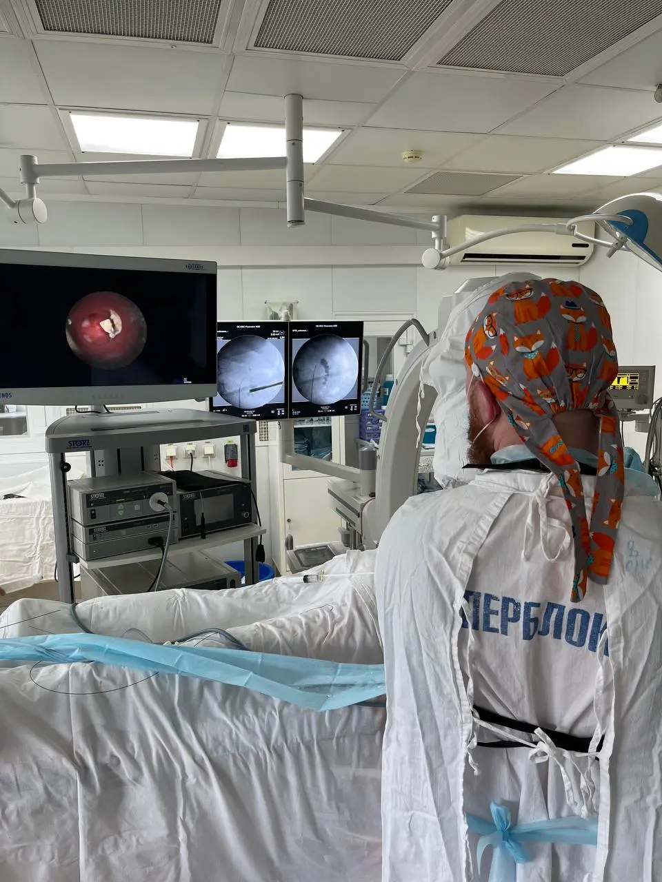
Percutaneous nephrolithotripsy: endoscopic and radiological monitoring combined in one operating position
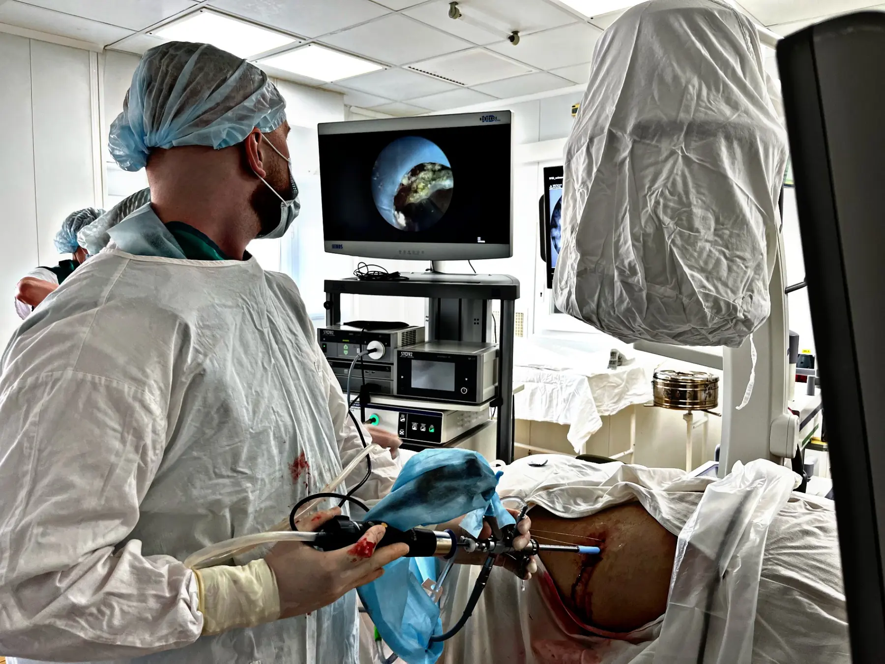
Percutaneous surgery with the patient supine: the endoscopic view and X-ray guidance are monitored simultaneously

The operating team at work during the endoscopic stage of the procedure

Visual monitoring of the endoscopic stage and instrument position
Percutaneous surgery for a staghorn stone
An excerpt from percutaneous surgery for a complex stone with the patient supine. The video has no sound; the decision to use a tubeless technique can be made only on the basis of the individual operation.
Supine PCNL at BMI 41
An excerpt from a percutaneous procedure in a patient with BMI 41. Patient position and access route are selected individually based on anatomy, anaesthetic factors and imaging.
Show additional materials7
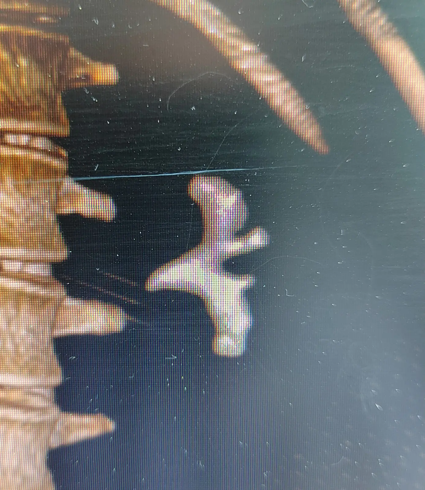
A staghorn stone occupies several parts of the renal collecting system; access is planned using the original CT data
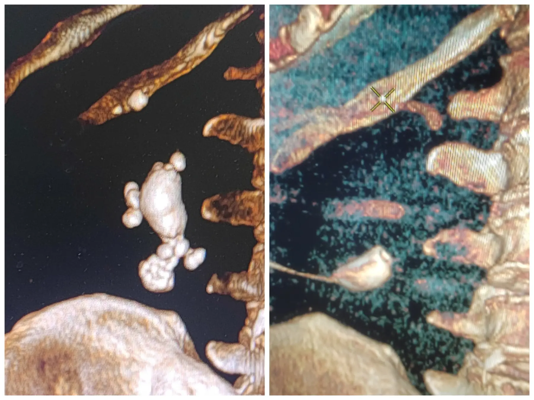
Comparison of CT findings before and after the procedure in one clinical case. An individual result is not a guarantee for another patient
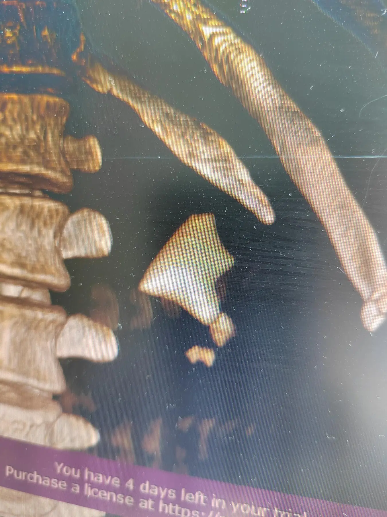
A complete staghorn stone on CT. These images are used to assess stone burden and plan access
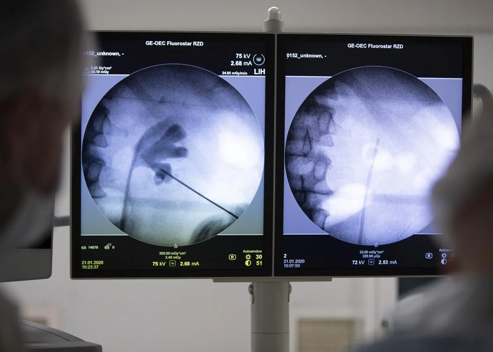
X-ray guidance helps control the direction of access to the kidney's collecting system
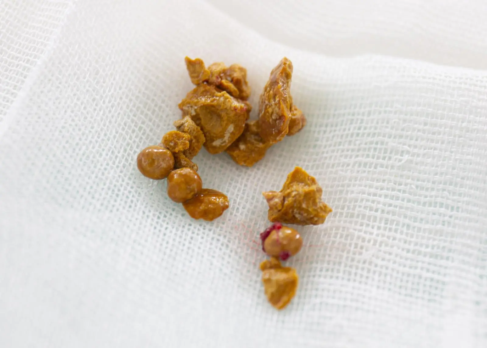
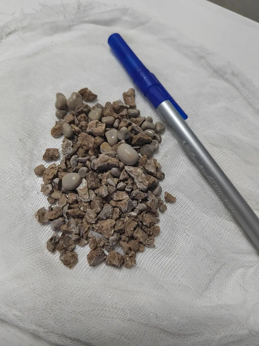
Removed fragments. Their appearance alone does not establish stone composition — laboratory analysis is required
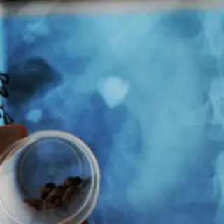
An archived image of fragments removed after a percutaneous procedure. The final outcome is assessed by follow-up imaging, rather than by a photograph
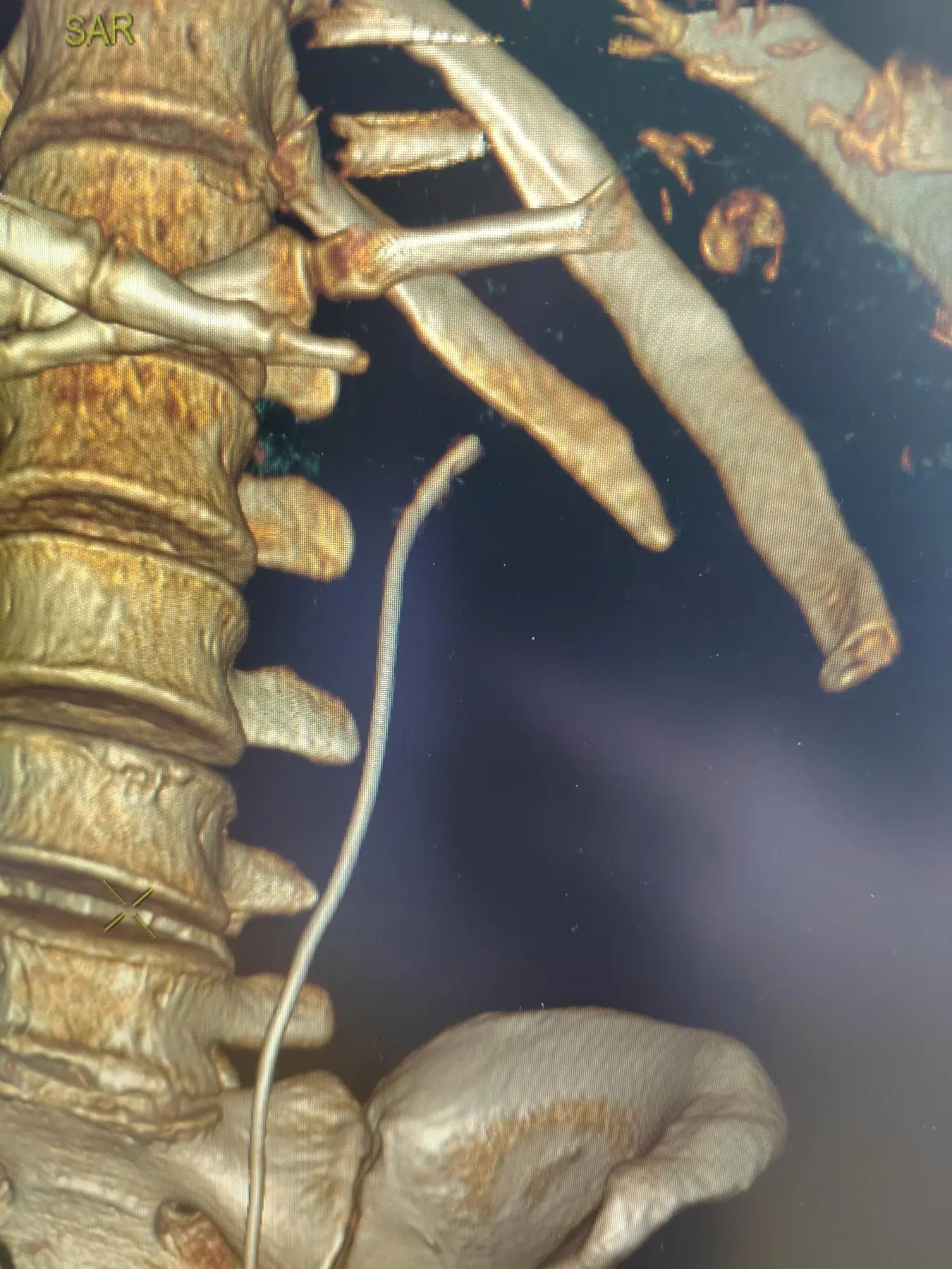
Follow-up CT on the next day in one clinical case. An individual result is not a promise of a similar result for another patient
04 / Preparation
What to prepare before a consultation
- Urinary tract CT in DICOM format and the report; contrast if indicated
- urinalysis and urine culture
- creatinine and kidney function measures
- complete blood count and coagulation tests
- discharge summaries from previous operations, and details of stents and nephrostomies
- a complete list of medications, allergies and other illnesses
05 / After surgery
Follow-up and recovery
After the procedure, temperature, blood tests, urine colour, drain function and urine drainage are monitored. The timing of nephrostomy or stent removal is determined individually.
Follow-up imaging shows whether clinically significant fragments remain. Stone composition analysis and metabolic assessment are important next steps: treatment is not complete after stone removal if recurrence prevention has not been addressed.
06 / Risks
What is discussed before the procedure
- bleeding and the possible need for additional measures to stop it
- infection, fever and sepsis
- injury to the urinary tract or neighbouring organs
- urine leakage and a temporary need for drainage
- residual fragments, an additional access tract or a further treatment stage
07 / Urgent care
When emergency care is needed
Fever or chills with impaired urinary drainage, no urine output, a sharp drop in urine output with a solitary kidney, uncontrolled pain or vomiting require calling 103/112 or going to the nearest urology hospital.
After surgery, a high temperature, increasing weakness, heavy bleeding with clots or cessation of drainage require urgent assessment.
Patient questions
Frequently asked questions
Can a staghorn stone be removed in one operation?
Sometimes, yes, but this cannot be guaranteed in advance. The possibility of a single stage depends on stone burden and location, kidney anatomy, procedure duration and the safety of additional access tracts.
Is a nephrostomy tube always left after PCNL?
No. The decision to place a nephrostomy tube or an internal stent is made based on the operation's findings. A tubeless technique is not suitable for every case.
What to bring to the appointment besides the CT report
Bring the actual CT images in DICOM format, urinalysis and urine culture, creatinine results, discharge summaries from previous procedures and a medication list. A written report alone is often insufficient for planning access.
Verifiable evidence
Sources
This page explains general guidance in plain language. Decisions for an individual patient are made after an in-person assessment and cannot be reduced to a single guideline recommendation.
In-person assessment and a clear plan