A clinical counterexample · strategy matters more than a template
Stones up to 55 mm — without a single incision
A surgeon's strength lies not only in the ability to operate. It also lies in knowing when to choose rather than surgery, when the evidence points to a safer approach.
01 / The system's verdict
The standard pathway posed an unacceptably high risk for this patient
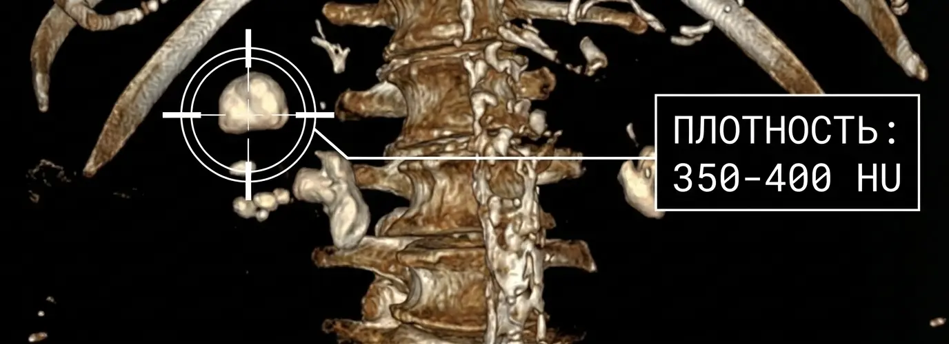
The patient was 73 years old. Multislice CT revealed bilateral staghorn stones of a critical size:
- right kidney — up to 40×55 mm, with moderate hydroureteronephrosis;
- left kidney — up to 30×42 mm;
- anaesthetic status — ASA IV.
Risk was determined not by age alone, but by severe comorbidity: class III obesity (BMI 41,9), coronary artery disease, post-infarction cardiosclerosis, type 2 diabetes mellitus, COPD and chronic heart failure.
For this stone burden, PCNL is usually the first-line treatment. However, in this case, the surgical and anaesthetic risk of elective PCNL was assessed as extremely high. The alternative was long-term nephrostomy drainage: external tubes, care and regular replacements without addressing the cause.
02 / A strategic crossroads
Three paths. Very different consequences of the decision
| Care pathway | What it offered | The cost of the decision |
|---|---|---|
| Gold standardPercutaneous surgery | Direct removal of large and staghorn stones | Extremely high risk life-threatening perioperative complications |
| Initial planNephrostomy drainage | Ensuring urinary drainage | Reduced quality of life: the stones remain, and the drains require care and replacement |
| Selected strategyControlled chemolysis | A chance to dissolve suspected uric acid stones without surgery | Complete dissolution in this case — with close monitoring and high treatment adherence |
03 / A change of paradigm
One CT parameter changed the entire treatment pathway
On re-evaluation of the multislice CT, stone density was only 350–400 HU, and the initial urine pH was 5,0. Together, these findings strongly suggested a uric acid composition.
This was not conclusive proof. However, the combined CT, radiographic and laboratory findings justified moving away from the standard approach and choosing intensive, closely monitored oral chemolysiswith a citrate mixture.
04 / Designing for adherence
A complex regimen was turned into four clear steps
A complex daily regimen in an older patient is more likely to lead to mistakes or treatment discontinuation. The care pathway was therefore designed around this patient's actual limitations.
Digital precision
Reduced vision made paper strips difficult to read. They were replaced with an electronic pH meter. Fresh urine pH was checked throughout the day and kept within the range prescribed by the doctor 6,8–7,2. Higher does not mean better: excessive alkalinisation increases the risk of other stone types.
Tolerance of the solution
The well-diluted citrate solution was taken through a straw to reduce contact with the front teeth and aversion to the taste, followed by rinsing the mouth with water. This is a supportive measure, not a guarantee of enamel protection.
Individualised night-time hydration
A glass of water and the evening dose of medication were taken before bed, with another glass during a usual night-time awakening. This shortened the prolonged acidic urine interval overnight. Because of heart failure, the total volume was not increased without medical advice — only within the individual limit.
Visual tracking
Each reading within the target range was marked in green on a large wall calendar. Contact with the doctor every two weeks helped maintain the routine and allowed timely adjustments to the regimen.
05 / Outcome after 4 months
Stone-free without surgery
On the follow-up multislice CT dated 19 January 2024 no stones were visible in either kidney, and urine drainage was unobstructed. This was complete dissolution in a single clinical case, not a universal guarantee.
- Uric acid
- 540→230µmol/L · normal blood uric acid
- Creatinine
- 123→109µmol/L
- Urea
- 9,9→7,5mmol/L
- Urine pH
- 5,0→6,8–7,0stable
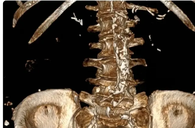 Before treatmentAfter 4 months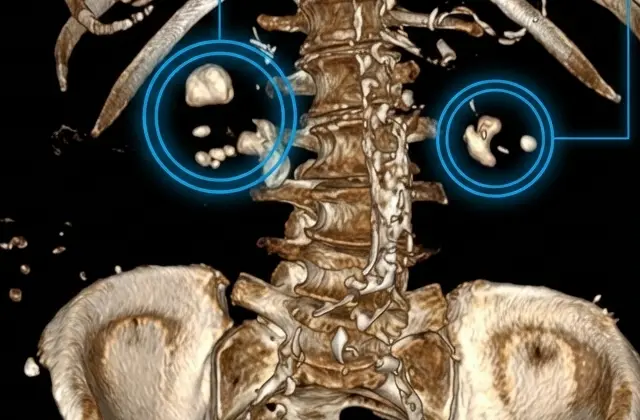
Verifiable evidence